
Keratoconus in Tijuana: early detection with corneal tomography

Keratoconus is one of the conditions I most enjoy explaining to my patients, because when it's caught early, there's a lot we can do. Here's what it is, when we should suspect it, and how I diagnose and treat it at Vision Clinic.
What is keratoconus?
Keratoconus is a progressive condition in which the cornea —the clear surface at the front of the eye— gradually thins and loses its normal spherical shape, taking on an irregular cone-like form. This causes irregular astigmatism and image distortion that, over time, can significantly affect vision. It usually begins in adolescence or early adulthood and progresses slowly over several years.
It's estimated to affect more than 23 million people worldwide, and detection rates have risen noticeably in recent years —not because there are more new cases, but because we now have technology capable of identifying it at much earlier stages than before.
When should we suspect it?
Keratoconus isn't always obvious on a standard eye exam. There are certain signs that should raise suspicion and warrant an evaluation with corneal tomography:
Very high or irregular astigmatism
Prescriptions with elevated astigmatism, especially if it's irregular or increasing from one exam to the next.
Frequent prescription changes
You need to update your prescription more often than usual because your vision keeps changing.
Children with high prescriptions
Significant prescriptions at a young age, when the eye normally shouldn't need that much correction yet.
Vision that doesn't reach 100% with glasses
Even with the correct prescription, visual acuity doesn't reach 100% with glasses (frame lenses).
Eye allergies and eye rubbing
People with allergies or who rub their eyes frequently, with blurry vision that isn't fully explained.
If you recognize yourself in any of these signs, I invite you to get a Sirius Plus corneal tomography at Vision Clinic —a quick, painless study that lets us see the true shape of your cornea with extremely high precision.
How do we diagnose it, and what treatment do I recommend?
In the same visit we have corneal tomography available, which lets me not only diagnose keratoconus, but also stage how advanced it is —key information for deciding on the most appropriate treatment for you.
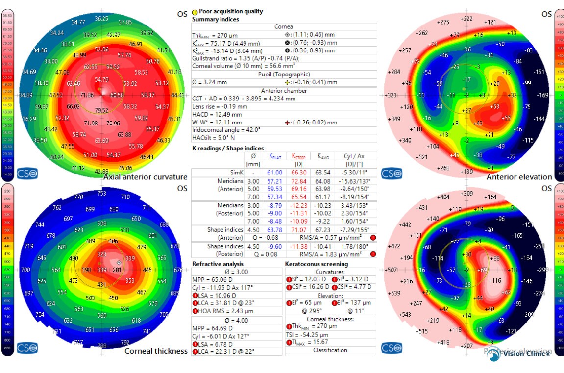
One point I want to make very clear: corneal crosslinking is, to date, the only treatment proven to stop the progression of keratoconus. It works by strengthening the collagen fibers in the cornea using ultraviolet light and vitamin B2 (riboflavin), and it's the foundation of treatment whenever we detect progression of the disease.
Beyond crosslinking, there are other therapies that don't stop the disease on their own, but do help improve your visual capacity and comfort with lenses:
- Intrastromal rings: segments placed inside the cornea to flatten and regularize its shape, improving vision and tolerance to contact lenses.
- CAIRS: a variant of the rings that uses corneal tissue instead of synthetic segments, with a similar goal.
- Athens protocol: combines crosslinking with topography-guided laser ablation to regularize the corneal surface and improve vision.
- Toric ICL: an implantable intraocular lens that corrects irregular astigmatism when contact lenses are no longer enough.
- Scleral contact lenses: larger-diameter rigid lenses that rest on the sclera and "vault" over the irregular area of the cornea, creating a regular optical surface and significantly improving vision without surgery.
As a cornea specialist, I'm the one who evaluates, diagnoses, and follows up with every keratoconus patient at Vision Clinic —from the first visit through every follow-up exam, to make sure the disease stays controlled and your vision, protected.